Sabado, Oktubre 1, 2011

Light Independent Reaction

The Light Independent Reaction

1.Carbon Fixation

            The cycle incorporates each CO2 molecules by attaching it to a five-carbon sugar RuBP (Ribulose Bisphosphate). The enzyme that catalyzes this step is called rubisco (RuBP carboxylase). The product of phase 1 is a 6 carbon sugar so unstable that it splits in half forming two molecules of 3-phosphoglycerate.


2. Reduction

            Each moleculeof3-phosphoglycerate
receives an additional phosphate group from ATP, becoming 1,3·bisphosphoglycerate. Next, a pair of electrons donated from NADPH reduces 1,3-bisphosphoglycerate, which also loses a phosphate group, becoming G3P. Specifically, the electrons from NADPH reduce a carboxyl group on 1,3-bisphosphoglycerate to the aldehyde group of G3P, which stores more potential energy. G3P is a sugar-the same three-carbon sugar formed in glycolysis by the splitting of glucose Notice that for every three molecules of CO2 that enter the cycle, there are six molecules ofG3P formed. But only one molecule of this three-carbon sugar can be counted as a net gain of carbohydrate. The cycle began with 15 carbons' worth of carbohydrate in the form of three molecules of the five-carbon sugar RuBP. Now there are 18 carbons' worth of carbohydrate in the form of six molecules of G3P. One molecule exits the cycle to be used by the plant cell, but the other five molecules must be recycled to regenerate the three molecules of RuBP.

3. Regeneration

            In a complex series of reactions. the carbon skeletons of five molecules of G3P are rearranged by the last steps of the Calvin cycle into three molecules of RuBP. To accomplish this, the cycle spends three more molecules of ATP. The RuBP is now prepared to receive CO2 again, and the cycle
continues. For the net synthesis of one G3P molecule, the Calvin cycle consumes a total of nine molecules of ATP and six molecules of NADPH. The light reactions regenerate the ATP and NADPH. The G3P spun off from the Calvin cycle becomes the starting material for metabolic pathways that synthesize other organic compounds, including glucose and other carbohydrates. Neither the light reactions nor the Calvin cycle alone can make sugar from CO2, Photosynthesis is an emergent property of the intact chloroplast, which integrates the two stages of photosynthesis.


Light Dependent Reactions

A. Linear Electron Flow
1. Light or photon strikes the leaf and the thylakoid capture it. As it enter the thylakoid it will first undergo a process in the Photosystem II. It will carry the the 2 atoms of hydrogen splitted from water(H2O)

2. The electron “bounce and bounce” in the pigment, as it hit the Chlorophyll A(P680), this will put the electron to the primary electron acceptor.

3. The photoexcited electron will be transferred by the Primacy Electron Acceptor to the photosystem 1 through the Electron Transport Chain (ETC). The electron will cross the ETC through its three parts; first the plastoquinone, after here to the cytochrome complex, the biggest membrane in ETC, in the CC is the ATP mill. The ATP mill pushes a phosphate to ADP present there becoming ATP as the electron roll over. After here it will pass the plastocyanine and at last to the Photosystem 1.

4. In the photosystem 1; the electron is in its ground-state level to ‘excite’ it, another electron enter through and the Chlorophyll A (P700) will again carry the electron to the primary electron acceptor. In both sides of the PEC here, Ferredoxin is situated. Fd is a protein that carries electron to the NADP+ reductase. In here hydrogen is added to NADP+  resulting in the formation of NADPH+H+.


B. Cyclic Electron Flow
1. Light or photon strikes the leaf and the thylakoid capture it. As it enter the thylakoid it will first undergo a process in the Photosystem II. It will carry the the 2 atoms of hydrogen splitted from water(H2O)

2. The electron “bounce and bounce” in the pigment, as it hit the Chlorophyll A(P680), this will put the electron to the primary electron acceptor.

3. The photoexcited electron will be transferred by the Primacy Electron Acceptor to the photosystem 1 through the Electron Transport Chain (ETC). The electron will cross the ETC through its three parts; first the plastoquinone, after here to the cytochrome complex, the biggest membrane in ETC, in the CC is the ATP mill. The ATP mill pushes a phosphate to ADP present there becoming ATP as the electron roll over. After here it will pass the plastocyanine and at last to the Photosystem 1.

4. The electron will be in the ground-state level in the P700 (Chlorophyll A), and a photon will enter PS1 ‘exciting’ the electron again. After that Chlorophyll A will transfer the electron to the Primary Protein Acceptor, in here Ferredoxin will carry the electron back to the Cytochrome Complex. In there it will again roll over the ATP mill. As it did so, ATP is generated by the addition of phosphate to ADP. And the cycle continues. 


Photosynthesis

What  is PHOTOSYNTHESIS?
Photosynthesis is the very nature of plants to produce their own food by converting light energy from the sun to chemical energy in the form of glucose. It occurs in the chloroplast; a part of the leaf that can be found in the mesophyll layer.

Light Dependent Reaction
Light Dependent Reaction is a process in which photosynthesis needs light to work out. In the thylakoid, a part in the chlorophyll, there are two photosystems that are involved in the process. The photosystem II and  photosystem I. They were named in order of discovery but photosystem II functions first in the light reactions.

  1. Photosystem II- P680 can absorb light efficiently within 680 nm.
  2. Photosystem I- P700 can absorb light efficiently within 700 nm.
Processes in Light Dependent Reaction
  1. Linear electron flow
  2. Cyclic electron flow
Light Independent Reaction
Light Independent Reaction is a process of undergoing photosynthesis without the presence of light. It is also known as the Calvin-Benson Cycle because it was discovered by Melvin Calvin and Andrew Benson. It is an anabolic process, building carbohydrates from smaller molecules and consuming energy. It produces a carbohydrate called glyceraldehyde-3-phosphate

*In the next post, you will learn more about light dependent & independent reactions.

Anatomy of a Leaf

The Anatomy of a Leaf
Outer Structure
  1. midrib - the central rib of a leaf - it is usually continuous with the petiole.
  2. petiole - a leaf stalk; it attaches the leaf to the plant.
  3. stipule - the small, paired appendages (sometimes leaf-life) that are found at the base of the petiole of leaves of many flowering plants.
  4. vein (vascular bundle) or small netted vein - Veins provide support for the leaf and transport both water and minerals (via xylem) and food energy (via phloem) through the leaf and on to the rest of the plant.
  5. blade - the outline of the leaf
Inner Structure
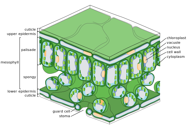
  1. cuticle - the waxy, water-repelling layer on the outer surface of a leaf that helps keep it from dying out (and protect it from invading bacteria, insects, and fungi). The cuticle is secreted by the epidermis (including the guard cells) and is often thinner on the underside of leaves. The cuticle is generally thicker on plants that live in dry environments.
  2. epidermis - the protective, outler layer of cells on the surface of a leaf. The guard cells (and stoma) are part of the epidermis. The surface of many leaves is coated with a waxy cuticle which is secreted by the epidermis.
  3. guard cell - one of a pair of sausage-shaped cells that surround a stoma (a pore in a leaf). Guard cells change shape (as light and humidity change), causing the stoma to open and close.
  4. mesophyll - the chlorophyll-containing leaf tissue located between the upper and lower epidermis. These cells convert sunlight into usable chemical energy for the plant.
  5. palisade mesophyll - a layer of elongated cells located under the upper epidermis. These cells contain most of the leaf's chlorophyll, converting sunlight into usable chemical energy for the plant.
  6. spongy mesophyll - the layer below the palisade mesophyll; it has irregularly-shaped cells with many air spaces between the cells. These cells contain some chlorophyll. The spongy mesophyll cells communicate with the guard cells (stomata), causing them to open or close, depending on the concentration of gases.
  7. stoma - (plural stomata) a pore (or opening) in a plant's leaves where water vapor and other gases leave and enter the plant. Stomata are formed by two guard cells that regulate the opening and closing of the pore. Generally, many more stomata are on the bottom of a leaf than on the top.


Leaf

What is a LEAF?
A leaf is responsible for:
  • photsynthesis
  • food production
  • transforming light energy to chemical energy
  • cellular respiration
Classifications of Leaf
A. Phyllotaxy - the fundamental study of leaf arrangement.
  1. Opposite - 2 leaves per node
  2. Alternate - one leaf per node
  3. Whorled - multiple leaves surrounding the node
  4. Spiral - leaves swirl around the plant.
*Node - attachment site of the leaf

B. Number of Cotyledon
  1. Monocot - has one cotyledon. It has a parallel vein leaf and scattered vascular vessel
  2. Dicot - has two cotyledons. It has a netted vein leaf and ring form vascular vessel

Monocot: left, parallel vein leaf; right, scattered vascular vessel
Dicot: left, netted vein leaf; right, ring form vascular vessel
  • Vascular Tissues are the transport vessels of plants
  • Xylem -upward; water and minerals
  • Phloem -downward; nutrients


Membrane Transport

Membrane Transport include the following:
  1. Passive Transport
  2. Active Transport
  3. Exocytosis
  4. Endocytosis
  5. Phagosytosis
  6. Pinocytosis
What is a PASSIVE TRANSPORT?
Passive transport is a diffusion of a substance across a membrane with no energy investment. It is the tendency of molecules of any substance to spread out evenly in space. It is driven by intrinsic kinetic energy of molecules. A molecule or ion that causes a membrane by moving down a concentration of electrochemical gradient and without expenditure of metabolic energy is said to be transported passively.

  • Simple diffusion - higher concentration to lower concentration
  • Facilitated Diffusion - molecules can go inside the cell membrane by transport protein
  • Osmosis - transport of water or gradient through a semi-permeable membrane.

What is an ACTIVE TRANSPORT?
Active transport enables a cell to maintain its internal concentrations of small molecules that would otherwise diffuse across the membrane. It is performed by specific proteins embedded in the membranes. ATP supplies the energy for most active transport. It requires the cell to expend metabolic energy. 

What is EXOCYTOSIS?
Exocytosis takes place when a transport vesicle budded from the golgi apparatus moved by the cytoskeleton to the plasma membrane.

What is ENDOCYTOSIS?
Endocytosis takes place when a small area of the plasma membrane sinks inward to form a pocket.

What is PHAGOCYTOSIS?
Phagosytosis is also known as the "cellular eating" process. The cell engulfs a particle by extending pseudopodia around it in a large vesicle.

What is PINOCYTOSIS?
Pinocytosis is also known as the "cellular drinking" process and is the counterpart of phagocytosis. A cell creates a vesicle around a droplet of extracellular fluid. All included solutes are taken in into the cell in this non-specific process.

Macromolecules

What are MACROMOLECULES?
Macromolecules are molecules needed to survive basic reactions.

Terms that you need to know:

  1. Polymers are long molecule consisting of many similar or identical building blocks linked by covalent bonds
  2. Dehydration Reaction is the loss of a water molecule to make a bond.
  3. Hydrolysis is the addition of a water molecule to break a bond
The Different Macromolecules
  • Carbohydrates are composed of hydrogen, carbon, and oxygen. This is the energy used by our body. The building block of carbohydrates is glucose. There are three types of carbohydrates:
1. Monosaccharides are the simplest form of sugar. Examples are glucose, fructose, and galactose.
2. Disaccharides are 2 monosaccharides joined by glycosidic linkage
  • Glucose + Glucose = Maltose
  • Glucose + Fructose = Sucrose
  • Galactose + Glucose = Lactose
  • Galactose + Fructose = Lactulose 
3. Polysaccharides are the complex of carbohydrates. An example is the chitin which is the carbohydrate used by arthropods to build their exoskeleton.

  • Fats make up the cellular membrane. Fats are the stored energy of our body. They are cushion that protect the internal organs from external force. The building block of fats are fatty acids.
  1. Fatty Acid is a long carbon skeleton, usually 16 or 18 carbon atoms in length.
  2. Phospholipids are lipids whose head is glycerol (hydrophilic) and tail is fatty acid (hydrophobic) which can be found on the triacylglycerol.
  3. Ester linkage is the link of the glycerol to the fatty acid.
  4. Saturated fats are animal fats which are solid in form.
  5. Unsaturated fats are plant fats which are in liquid form and healthy fats. It has a kink which makes it more stable.
  6. Steroids are mistakenly known as proteins that help build muscles. Steroids are fats that strengthen the muscles.
  • Nucleic Acid is the basic composition of DNA and RNA.

Location
# of strands
Nitrogenous bases
DNA
Nucleus
2
C, G, A,T
RNA
In & out of the nucleus
1
C, G, A, U

  1. Nucleotide is more stable and the building block of nucleic acid. 
  2. Nucleoside is like a nucleotide except that it has no phosphate. It is the building block of nucleotide.
  • Protein is made up of amino acids and serves as the building blocks of cells.
  • Enzymes are specific proteins that mostly end up in -ase


20 AMINO ACIDS


Name
Abb
Name
Abb
Name
Abb
Name
Abb
Name
Abb
Glycine
Gly
Isoleucine
Ile
Proline
Pro
Tyrosine
Tyr
Glutamic Acid
Glu
Alanine
Ala
Methionine
Met
Serine
Ser
Asparagine
Asn
Lysine
Lys
Valine
Val
Phenylalanine
Phe
Threonine
Thr
Glutamine
Gln
Arginine
Arg
Leucine
Leu
Tryptophan
Trp
Cysteine
Cys
Aspartic Acid
Asp
Histidine
His

  1. Primary Structure is where amino acids and polypeptide chains are formed.
  2. Secondary Structure is where coiled protein (alpha helix) and folded protein (beta-pleated sheet) are formed.
  3. Tertiary Structure is where interaction of molecules within the protein structure occur.
  4. Quaternary Structure is the final stage in which the protein knows its function.
The Protein Synthesis

Replication
Transcription
Translation
Location
Nucleus
Nucleus
Cytoplasm
Key player
DNA is copied
DNA is transcribed to RNA (codon)
RNA
(anti-codon=amino acid)


Huwebes, Setyembre 29, 2011

Functional Groups

What are FUNCTIONAL GROUPS?
Functional Groups are groups of specific atoms that are responsible for chemical reactions of molecules.

The Functional Groups you should know

  1. Hydroxyl is an alcohol group that dissolves organic bacteria or compound. Mostly, molecules that end in -ol belong to the hydroxyl group like alcohol, methanol, ethanol, etc.
  2. Carbonyl is a functional group in which a carbon atom is joined to an oxygen atom by double bond. It gives sugar molecules longer polymers especially in carbohydrates. There are two carbonyl groups: (1) Ketose in which the carbonyl group is within the carbon skeleton; and (2) Aldose in which the carbonyl group is at the end of the carbon skeleton. 
  3. Carboxyl is the functional group that is the combination of the hydroxyl and carbonyl group. In this functional group, an oxygen atom is double bonded to a carbon molecule that is bonded to a hydroxyl group. It is responsible for the formation of acid. 
  4. Amino is the functional group that is responsible for the creation of the base or the polypeptide chain. It is also the building block of protein.
  5. Sulfhydryl is the functional group that consists of a sulfur atom bonded to an atom of hydrogen, resembles a hydroxyl group in shape. Two sulfhydryl groups can react, forming a covalent bond. This “cross-linking” helps stabilize protein structure. Cross-linking of cysteines in hair proteins maintains the curliness or straightness of hair. Straight hair can be “permanently” curled by shaping it around curlers, then breaking and re-forming the crosslinking bonds.
The carbon skeleton of the five functional groups.

Introduction to Functional Groups

TERMS:
  1. Hydrogen bond is the weakest bond because oxygen is easily attracted.
  2. Carbon skeleton is the skeletal formula of an organic compound.
  3. Organic compound are molecules with the element of carbon.
  4. Isomer is the same number of molecules but has different molecular structure.
  5. Isomerization is the chemical process in which a compound is transformed into any of its isomeric forms.
Variations of Carbon Skeleton
  1. Length
  2. Branching - carbon on top of another carbon
  3. Double bond - represented by two connecting lines in a carbon skeleton
  4. Ring type - carbons form a ring
Types of Isomer
  1. Structural isomers are isomers with the same molecular formula but has different structure
  2. Geometric isomers are isomers with the same molecular formula but differ in spacial arrangement
  3. Enantiomer are isomers which are mirror images.

Osmosis and Tonicity in Cells

What is OSMOSIS?
OSMOSIS is the movement of water through a semi-permeable membrane.

What is TONICITY?
TONICITY is the study of water and solute. It is also the ability of the solution to lose or gain water.

Types of Tonicity in Cells
  1. Isotonic solution is the solution in which the amount of solute and solvent are equal. Animal cells that are submerged into an isotonic solution is normal while a plant cell will be flaccid or soft and hanging loosely. 
  2. Hypertonic solution is the solution wherein there is more solute than solvent. Animal cells in this solution tends to shrink and plant cells will undergo plasmolysis and will be plasmolyzed.
  3. Hypotonic solution is the solution in which there are more solvent than solute. Animal cells that are exposed to this solution will undergo lysis and will be lysed while a plant cell will be in its normal state. The reason why plants will be in normal in a hypotonic solution is because plants need more water. 
Image of the three types of tonicity in cells


Miyerkules, Setyembre 28, 2011

Plant Cells

What are PLANT cells?
Plant cells are cells that can obviously be found on plants. These are specialized cells that are appropriate for storing large amounts of water for food production, respiration, and other processes. These cells can also capture light from the sun to be used in making food.

Parts of a Plant cell
  1. Cell wall is the outer layer that maintains the cell's shape and protects the cell from mechanical damage; made of cellulose, other polysaccharides and protein
  2. Plasmodesmata channels through cell walls that connect the cytoplasms of adjacent cells.
  3. Chloroplast is a photosynthetic organelle. It converts energy of sunlight to chemical energy stored in sugar molecules.
  4. Central Vacuole is a prominent organelle in older plant cells. Its functions include storage, breakdown of waste products, hydrolysis of macromolecules, enlargement of vacuole is a major mechanism of plant growth
  5. Nucleus
  6. Rough Endoplasmic Reticulum
  7. Smooth Endoplasmic Reticulum
  8. Ribosome
  9. Golgi Apparatus
  10. Mitochondrion
  11. Peroxisome
  12. Plasma Membrane
  13. Cytoskeleton
Some notes:
To add some information on chloroplasts:
Chloroplasts contain cholorphyll that capture, absorb, and reflect light for food production.
Chlorophyll A is the chlorophyll which reflects the green pigment we commonly see in plants.
Chlorophyll B is the chlorophyll which reflects the yellow green pigment.
Chlorophyll C is the chlorophyll which is an accessory pigment which reflects any pigment.
Carotenoids reflect the orange color like we see on carrots.


Martes, Setyembre 27, 2011

Animal Cells

Animal cells are the cells that can be found in the animals and the human body. A group of cells form tissues.

Parts of the Animal cell

  1. Flagellum are the locomotion organelles present in some animal cells; composed of a duster of microtubules within an extension of the plasma mebrane
  2. Centrosome is the region where the cell's microtubules are intiated; contains a pair of centrioles 
  3. Microvilli are projections that increase the cell's surface area
  4. Cytoskeleton reinforces the cell's shape; cellular locomotion; components are made up of protein. This includes the Microtubules, Microfilaments, and Intermediate filaments.
  5. Mitochondrion is the organelle where ATP is produced and where cellular respiration occurs.
  6. Peroxisome is the organelle with various specialized metabolic functions; produces hydrogen peroxide as a by-product and converts it to water
  7. Lysosome is a digestive organelle where macromolecules are hydrolyzed
  8. Golgi Apparatus is the organelle active in synthesis, modification, sorting, and secretion of cell products
  9. Ribosomes are organelles that are responsible for protein poduction
  10. Cell Membrane/Plasma Membrane is the membrane that encloses the cell and protects it from unwanted substances. This also regulates what goes in and out of the cell.
  11. Endoplasmic reticulum is the network of membranous sacs that is active in synthesis and has rough and smooth regions according to the ribosomes studded on them.
  12. Nucleus- contain the DNA and genetic information of the cell.
  • Nuclear envelope is the double membrane enclosing the nucleus
  • Nucleolus is the structure involved in the production of ribosomes
  • Chromatin is the material consisting of DNA and proteins


Eukaryotic Cells

What are EUKARYOTIC CELLS?
Eukaryotic Cells are cells that contain a nucleus and membrane-bounded organelles. Its name is derived from the Greek words "euka" and "karyose" which means true nucleus.

Types of eukaryotic cells
There are two types of eukaryotic cells. A type is an animal cell and the other is the plant cell.

Animal cells
Animal cell is a type of a eukaryotic cell that makes up many tissues in animals.
There are many different cell types. According to a study, there are approximately 210 distinct cell types in the adult human body.

Plant cells
Plant cell is a type of a eukaryotic cell that are only found in plants.
This cell have special characteristics that suit the needs of the plants like a large water storage and a site for their food production.

Archaea

What are Archea?
Archaea are prokaryotes that live in extreme places. They may live in places that are extremely hot, cold, acidic, etc. With this discovery, archaea are also called extremophiles.

Classifications of Archaea

  1. Halophiles are archaea that live in extremely salty areas.
  2. Acidophiles are archaea that live in extremely acidic places.
  3. Thermophiles are archaea that live in extremely hot areas.
  4. Methanogens are archaea that are found in areas with methane.
  5. Psychrophiles are archaea that live in extremely cold places.
  6. Alkaliphiles are archaea that thrive in alkaline environments with a pH of 9 to 11.
Halophiles

Acidophiles

Thermophiles

Methanogens


Psychrophiles

Alkaliphiles

Lunes, Setyembre 26, 2011

Bacteria

Bacteria-these are the single celled organisms that are found everywhere.
Types of Bacteria:
  • According to SHAPE:
  1. cocci-sphere shaped bacteria
  2. Streptococci bacteria
  3. bacilli-rod shaped bacteria
  4. Lactobacilli bacteria
  5. spirilla-spiral shaped bacteria


Spirillum bacteria
  • According to AMOUNT OF PEPTIDOGLYCAN present
  1. Gram-positive-It contains more peptidoglycan and has a less complicated structure
  2. A gram-positive bacteria that is stained violet
  3. Gram-negative-It contains less peptidoglycan and has a more complicated structure
A gram-negative bacteria that is stained red
  • According to OXYGEN REQUIREMENT
  1. Aerobic-requires oxygen for them to survive
  2. Aerobic bacteria
  3. Anaerobic-does not need oxygen for survival
  4. Anaerobic bacteria
  5. Facultative-are bacteria that may live with or without oxygen to survive
Facultative bacteria
  • Heterotrophic and Autotrophic bacteria
  1. Autotrophic-obtain the carbon it needs from carbon dioxide
  2. Heterotrophic-obtain carbon and/or sugar from their surroundings